

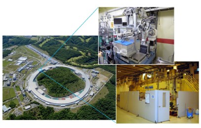
Beamline for Biological Macromolecular Assemblies at SPring-8 (BL44XU)
【Year established】 1999 (Upgraded in 2001, 2003, 2006, 2009, 2011, 2012, 2015, 2018)
【Facility specification】
Light source: Undulator
Optics: N2-cooled double crystal monochromator, Horizontal and vertical focusing mirrors
Detector: Pixel array detector (Eiger 16M)
【Description】
The beamline for X-ray crystallography of biological macromolecules at SPring-8 (BL44XU) is available to researchers who belong to academic organizations throughout the world. Regular applications involving non-proprietary proposals are received once per year, and urgent applications can be accepted all year around. More than 50% of the total beamtime is used for collaborative research programs of the Institute for Protein Research. This beamline is specially designed for data collection of large unit cell crystals for analyzing biological macromolecular assemblies, such as protein complexes, protein-nucleic acid complexes, and viruses.
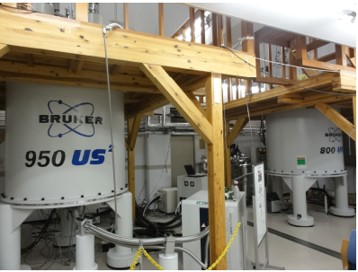
Solution NMR Spectrometer in an Ultra-high Magnetic Field
【Year established】 2010 (Upgraded in 2013 and 2014)
【Equipments】
950 and 800 MHz NMR Spectroscopy (BRUKER)
Spectrometer : Avance III 950, Avance III HD 800 (2013 upgraded)
Probe : Triple resonance cryogenic probe (1H,13C,15N)
【Description】
The ultra high field NMR instruments ( ¹H resonance frequency of 950 and 800 MHz) enable the analysis of 3D structures dynamics and interactions of high molecular-weight protein complexes and organic molecules at low concentrations, which markedly exceeds the performance of conventional spectrometers. This device provides one of the world’s highest magnetic fields, following a 1000 MHz spectrometer in France.
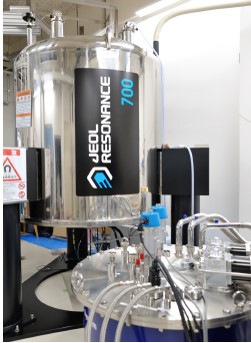
Solid-state NMR Spectrometers
【Year established】 2001 (Upgraded in 2006 and 2013)
【Equipment】
500MHz, 700MHz Solid-State NMR Spectrometers
600MHz/396GHz Solid-State DNP-NMR Spectrometer
700MHz/461GHz Solid-State DNP-NMR Spectrometer
【Description】
These spectrometers enable the analysis of the structures and functions of biomolecules in non-crystalline solid states, such as proteins in lipid bilayers and amyloid proteins. The two DNP-NMR spectrometers are equipped with gyrotrons generating high-power submillimeter waves and sample-rotation systems at cryogenic temperatures. Thereby, they generate hyper-polarization that provide the world’s highest performance in NMR sensitivity.
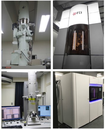
Cryo-electron Microscopes
【Year extablished】 2012 (Upgraded in 2015 and 2017)
【Equipments】
120kV H-7650(2010-)
200kV JEM-2200FS(2012-)
300kV Titan Krios G2(2015-)
200kV Talos Arctica(2017-)
【Description】
Cryoelectron microscopy (Cryo-EM) method has become powerful technology to determine 3D structure of macromolecules and received the Novel Prize in Chemistry 2017. IPR has an excellent lineup of cyro-EM machines and related equipments, enabling high-performance and high-throughput analyses. Three main machines are all equipped with direct electron detector (DED), and we have achieved determination of 2.3Å resolution structure in early 2018.
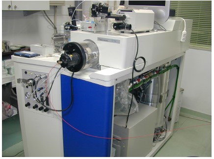
Analytical Apparatus for Supra-biomolecules
【Year extablished】 1999
【Equipments】
Q-TOF II (MS/MS)
【Description】
Knowledge of the chemical structures of biomolecules is indispensable for understanding their biological functions and roles. However, since they are normally obtained in limited amounts and consisting of various molecules with diverse chemical properties, their structures are difficult to analyze with conventional methods. The analytical apparatus for supra-biomolecules is a tandem mass spectrometer (MS/MS) equipped with the electrospray ionization method. It allows the detection of biomolecules at sub-picomole levels, especially proteins, peptides, and sugar chains of glycoproteins, and is used for their structural analyses.

High-performance X-ray Diffractometer
【Year established】 2003 (Upgraded in 2011 and 2016)
【Equipments】
X-ray generator: RIGAKU FR-E SuperBrightTM, Confocal VariMax Mirror
Detector: RIGAKU XtaLAB SynergyCustom/R-axis IV++
【Description】
This system has a state-of-the-art pixel array detector (HyPix600HE) and a high-performance image plate detector (R-AXIS VII) combined with a high-intensity rotating-anode X-ray generator (Rigaku FR-E) and confocal mirrors (Rigaku VariMax HF and CMF-MicroMax). This system gives a top-level performance X-ray diffraction data collection system for protein crystallography.