This page is for tutorial data and documents for education of electron microscopy single particle analysis.
A single particle analysis requires a huge amount of 2D image data to reconstruct a high-resolution 3D map. A typical 2D image size ranges from 100GB to 10 TB. These huge data are not good for educational purposes, because they require a long time to be downloaded and computer-processed. For educational purposes, we start to provide small 2D image data by choosing fine images from the EMPIAR database. We also prepare documents describing computer procedures for analyzing these data using the standard software, such as Relion.
These data and documents are free to use. We will be happy if they contribute to educate young EM researchers.
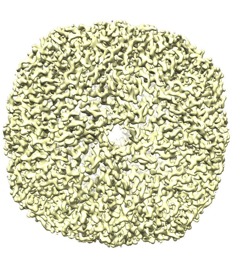
2D micrographs of apo ferritin taken by JEOL CRYO ARM 300 provides a 3D map with 1.54 angstrom resolution. However, the file size of the original data (EMPIAR-10248) is 145.9 G byte. We made a 1/100 tutorial data (1.7 G byte) [EMPIAR-10248_tutorial_500frames.tar] by choosing fine 2D images from the original images. This tutorial data is easy to be downloaded and to be processed, and quickly yields a 3D map with 2.85 angstrom.
| Original data | Tutorial data | |
| EMPIAR-10248 |
Images taken from
EMPIAR-10248. [EMPIAR-10248_tutorial_500frames.tar] |
|
| File size | 145.9 Gbyte | 1.7 Gbyte |
| Number of micrographs | 50 frames x 971 files | 25 frames x 20 files |
| 3D map | EMD-9865 | |
| Resolution (angstrom) | 1.54 | 2.84 |
 |
 |