JOINT USAGE / RESEARCH CENTERIPR Osaka University
The dead-line for submission:December 1st, 2022–
【Overview】 Institute for Protein Research (IPR), Osaka University, invites applications for international collaborative research. The research should be conducted in the form of a collaboration with at least one of the Principal Investigators (PIs) at IPR, or it should use equipments/ xperimental facilities of IPR, including solution- and solid-state-NMR spectrometers with ultra-high magnetic fields, cryoelectron microscopes, and a synchrotron beam line for biological macromolecular assemblies at SPring-8. In the fiscal year of 2023, the research period is from April 1st, 2023 to March 31st, 2024. Successful applications will receive financial support for the travel expenses, up to the amount determined at the time of acceptance based on the availability of the funds. Each applicant should submit “Application Form” for the collaborative research along with a recommendation from at least one PI at IPR (listed in the next page). The dead-line for submission for the 2023 fiscal year is December 1st, 2022. After review, accepted applications will be announced at the end of March, 2023. Even after the dead-line, proposals with urgent need can be submitted at any time during the 2023 fiscal year and may be approved after review, although the travel support may not be granted for such "late" proposals.
The dead-line for submission:December 1st, 2022–
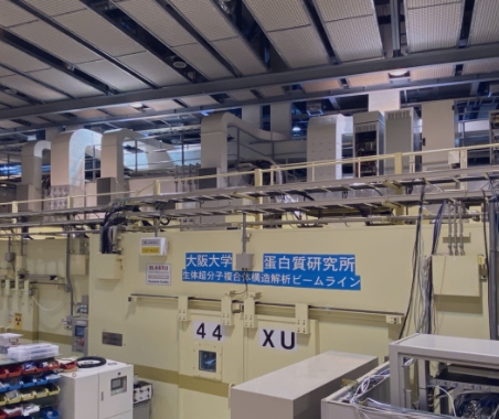
【Overview】 Institute for Protein Research is now accepting non-proprietary proposals for 2023 using the Beamline for Macromolecular Assemblies (SPring-8 BL44XU) operated by IPR. Beamline for Macromolecular Assemblies (SPring-8 BL44XU) This beamline (BL44XU) is specially designed for data collection of biological macromolecule assembly crystals, such as protein complexes, protein-nucleic acid complexes, and viruses. It uses an undulator as a light source. Detail of the beamline is shown in the web site. http://www.protein.osaka-u.ac.jp/rcsfp/supracryst/en/research/beamline/
The dead-line for submission:December 1st, 2022
Institute for Protein Research is now accepting non-proprietary proposals for Microcrystal Electron Diffraction (MicroED). Microcrystal Electron Diffraction (MicroED) MicroED (Microcrystal Electron Diffraction: Rotational method for the electron diffraction) is the crystallographic technique to collect the diffraction images from thin (< 1μm) 3D crystals in the powder sample. Even in the case that the X-ray experiment only showed the power diffraction pattern from the sample, MicroED experiment may acquire the diffraction patterns from single crystal(s) and solve the structure of complicated natural compounds or synthetic organic materials. Our institute implemented the automatic data acquisition system already, then crystal polymorph or low rate of crystalline particles may not be a serious problem. Since the method is still under development, applicants cannot operate the electron microscopy by themselves in principle and collaborative support from IPR member is essential.
We aim to establish a core center for basic and applied research on proteins where we elucidate their structures and functions, and can understand their biological roles from molecular to cellular and higher levels. This will lead to marked advancements in life sciences in Japan.
To achieve this, we promote domestic and international collaborations, provide opportunities to access various facilities and databases, and organize IPR seminars. Through these activities, we help stimulate communities studying life sciences on the basis of proteins.
Our activities as a Joint Usage/Research Center are guided by the Administrative Council and Panel on Join Usage/Research, members of which are selected from external persons with relevant knowledge and experience, so that we can respond adequately to community trends.
01
IPR offers the usage of its own synchrotron beamline at SPring-8, Nuclear Magnetic Resonance (NMR) spectrometers with ultra-high sensitivities and Cryo-electron microscopy at IPR, to domestic and international protein researchers.
02
IPR has developed a protein structural database (PDB: Protein Data Bank) as PDBj (PDB Japan), one of the four members of the wwPDB (worldwide PDB), by annotating the deposited data from structural biologists in Asia and Oceania and by providing several original services and derived databases. PDBj-BMRB is also constructing an NMR experimental database, collaborating with BMRB (BioMagResBank) in the U.S.A.
03
Through public invitations to join the 8 collaborative programs above, we contribute to domestic and international communities devoted to protein sciences and the development of human resources.
Our major activities are to promote 1. Joint usage of advanced facilities and equipment, 2. Access to databases, and 3. Collaborations including those leading to the development of human resources. Through these activities, we aim to activate protein and life science communities.
Domestic Collaborators
International Collaborative Research
SPring-8 Collaborative Research
NMR Collaborative Research
Cryo-EM Collaborative Research
MicroED Collaborative Research
IPR Seminars
IPR Fellows
The Joint Usage/Research Centers on the basis of organization and advanced facilities of IPR
Contribution to the communities studying life sciences on the basis of proteins
ⒸRIKEN
The state-of-the-art large experimental facilities housed in and operated by IPR cover all three major methodologies in the modern structural biology research field, namely, X-ray crystallography, nuclear magnetic resonance (NMR) spectroscopy, and cryo-electron microscopy (cryo-EM).
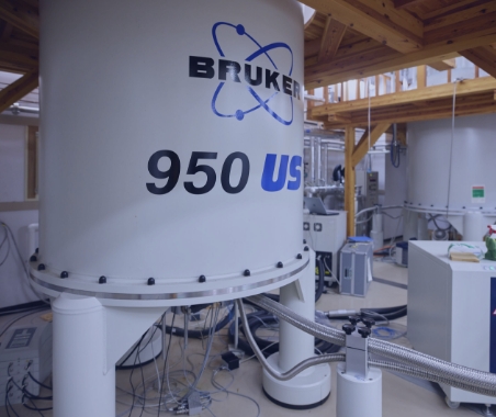
As for X-ray crystallography, we manage a contract beamline for macromolecular assemblies (BL44XU) at the SPring-8 synchrotron facility located in Nishi-Harima, Hyogo, ~120 km west from the institute. At the institute, as many as nine NMR spectrometers, including the flagship solution NMR model (950 MHz) and solid-state NMR equipped with an ultra-high sensitive DNP system, are housed and operated around the clock.
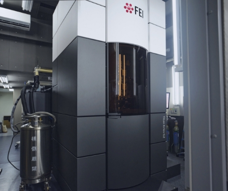
We also manage three cryo-EM scopes including Titan Krios (Thermo Fisher Scientific), Talos Arctica (Thermo Fisher Scientific), and JEM-2200FS (JEOL), all equipped with direct electron detector. They produce thousands of high-quality images every day to enable world-class single particle analyses for biologically important molecules.
Beamline for Biological Macromolecular Assemblies at SPring-8 (BL44XU)
The beamline for X-ray crystallography of biological macromolecules at SPring-8 (BL44XU) is available to researchers who belong to academic organizations throughout the world. Regular applications involving non-proprietary proposals are received once per year, and urgent applications can be accepted all year around. More than 50% of the total beamtime is used for collaborative research programs of the Institute for Protein Research. This beamline is specially designed for data collection of large unit cell crystals for analyzing biological macromolecular assemblies, such as protein complexes, protein-nucleic acid complexes, and viruses.
Year established |
1999 (Upgraded in 2001, 2003, 2006, 2009, 2011, 2012, 2015, 2018) |
---|---|
Facility specification |
Light source: |
Solution NMR Spectrometer in an Ultra-high Magnetic Field
The ultra high field NMR instruments ( ¹H resonance frequency of 950 and 800 MHz) enable the analysis of 3D structures dynamics and interactions of high molecular-weight protein complexes and organic molecules at low concentrations, which markedly exceeds the performance of conventional spectrometers. This device provides one of the world’s highest magnetic fields, following a 1000 MHz spectrometer in France.
Year established |
2010 (Upgraded in 2013 and 2014) |
---|---|
Equipments |
950 and 800 MHz NMR Spectroscopy (BRUKER)
Spectrometer: |
These spectrometers enable the analysis of the structures and functions of biomolecules in non-crystalline solid states, such as proteins in lipid bilayers and amyloid proteins. The two DNP-NMR spectrometers are equipped with gyrotrons generating high-power submillimeter waves and sample-rotation systems at cryogenic temperatures. Thereby, they generate hyper-polarization that provide the world’s highest performance in NMR sensitivity.
Year established |
2001 (Upgraded in 2006 and 2013) |
---|---|
Equipment |
500MHz, 700MHz Solid-State NMR Spectrometers |
Cryoelectron microscopy (Cryo-EM) method has become powerful technology to determine 3D structure of macromolecules and received the Novel Prize in Chemistry 2017. IPR has an excellent lineup of cyro-EM machines and related equipments, enabling high-performance and high-throughput analyses. Three main machines are all equipped with direct electron detector (DED), and we have achieved determination of 2.3Å resolution structure in early 2018.
Year extablished |
2012 (Upgraded in 2015 and 2017) |
---|---|
Equipments |
120kV H-7650(2010-) |
Analytical Apparatus for Supra-biomolecules
Knowledge of the chemical structures of biomolecules is indispensable for understanding their biological functions and roles. However, since they are normally obtained in limited amounts and consisting of various molecules with diverse chemical properties, their structures are difficult to analyze with conventional methods. The analytical apparatus for supra-biomolecules is a tandem mass spectrometer (MS/MS) equipped with the electrospray ionization method. It allows the detection of biomolecules at sub-picomole levels, especially proteins, peptides, and sugar chains of glycoproteins, and is used for their structural analyses.
Year extablished |
1991 |
---|---|
Equipments |
Q-TOF II (MS/MS) |
High-performance X-ray Diffractometer
Three X-ray diffractometers with cryo-stream coolers for protein crystallography are mounted on two rotating anode X-ray generators.
Year established |
2003 (Upgraded in 2011 and 2016) |
---|---|
Equipments |
X-ray generator: |
Amino acid sequence (primary structure) of a protein is an essential information for studying protein structure and function. Full-length amino acid sequences of proteins are easily obtained from the information of genome, however, proteins in the actual living system are sometimes truncated at N-terminus or/and C-terminus or modified by post-translational modification.
Therefore, chemical sequencing using Edman degradation method gives valuable sequence information. In addition, the amino acid sequences of proteins of a species without genome information must be directly analyzed with the protein samples themselves.
In the Institute for Protein Research (IPR), we have two protein sequencers (ABI Procise 491 cLC and Shimadzu PPSQ-53A) to analyze the N-terminal sequence of a protein or a peptide. We accept requests from outside IPR as commissioned analyses.
Year established |
2002 |
---|---|
Equipments |
Applied Applied Biosystems Procise 491 cLC |
Year established |
2019 |
---|---|
Equipments |
Shimadzu PPSQ-53A |
Institute for Protein Research manages the six world standard databases (CSD, PDB, BMRB, EMDB, EMPIAR and MOUSE BASEMENT MEMBRANE BODYMAP) as a public good, covering the wide range of molecular size from the small molecular organic compounds and protein molecules to the extracellular matrix, and containing diverse data, such as chemical spectra, atomic coordinates of molecular structure and micrographs. Our databases provide additional links to other experimental database, journals and archives of chemical compounds, which is regarded as a core data resource for supporting the fundamentals of frontier biosciences.
PDBj (Protein Data Bank Japan) maintains global structural databases of macromolecular structures (the PDB, BMRB and EMDB core archives) and provides integrated tools and services, in collaboration with RCSB PDB and BMRB in USA, and PDBe and EMDB in Europe.
Protein Data Bank (PDB) is a single global archive of the experimentally determined structures of biological macromolecules. The PDB core archive is maintained by the worldwide PDB consortium with four partners; the RCSB PDB and BMRB in USA, the PDBj in Japan, and the PDBe in UK. PDBj is a founding member of the wwPDB and currently processing all the depositions originated from Asia and middle-East. From Osaka University, PDBj is distributing all PDB data to the world (https://pdbj.org). (This activiy is supported by JST-NBDC and the MEXT grant of Joint Usage/Research allocated to IPR, Osaka University.)
The Biological Magnetic Resonance Data Bank (BMRB) is a repository for data from NMR Spectroscopy on Proteins, Peptides, Nucleic Acids, and other Biomolecules. The head office of BMRB is located in USA, but PDBj is operating a branch office of BMRB based on the academic exchange agreement. The BMRB branch office at PDBj (BMRBj) is accepting and processing all the data deposited to the server machines installed at PDBj. All released data are available from our BMRB server to the world. (This activity is supported by JST-NBDC)
The Electron Microscopy Data Bank (EMDB) is a public repository for electron microscopy density maps of macromolecular complexes and subcellular structures. The deposited data from Asia and middle-East are processed by PDBj, and all other regional entries processed by other partner sites are collaboratively released to the world. EMDB entries with low resolution does not accompany the atomic coordinates, but those with high resolution may accompany the atomic coordinates, which is stored in the PDB archive. (This activity is supported by AMED-BINDS)
The Electron Microscopy Public Image Archive (EMPIAR) is a public resource for raw, 2D electron microscopy images. The office of EMPIAR is operated by European Bioinformatics Institute (EBI) in UK. PDBj started the branch office activity of distributing and brokering the EMPIAR data from 2017, collaborating with EBI. We are planning more activities, such as curating and processing the regional data, in our branch office in near future. (This activity is supported by AMED-BINDS)
The Cambridge Structural Database (CSD) is one of the first numerical scientific databases in the world containing crystal structure data for all small molecules studied by X-ray or Neutron diffraction. The data have been archived from 1965 by the Cambridge Crystallographic Data Centre (CCDC) with continuing help from the crystallographic community worldwide. The first releases of the CSD System to the USA, Italy and Japan occurred in 1970’s and the Institute for Protein Research was in charge of distribution in Japan. Now, the CSD System is made available to academic users through the National Affiliated Centres (NACs), who undertake licensing and distribution of the media on the CCDC’s behalf in a specific country. Institute for Protein Research, Osaka University, is the NAC in Japan.
Many cell types in our body sustain their functions through adhesive interactions with a sheet-like extracellular matrix called “basement membrane”. This database compiles high-resolution immunohistochemical images for a total of 42 basement membrane proteins including all subunit chains of laminins, collagen IV, and heparan sulfate proteoglycans (perlecan, agrin, and collagen XVIII) in mouse embryos. One can move around on a virtual slide of a whole mouse embryo and magnify any part of the embryo to locate basement membrane proteins of interest at a single cell level.
The research results have to be reported within one month after completion of the collaborative research.
Please submit “2022 Summary of Research Report Form” to us by May 12, 2022.
*The forms are written in English.
*”Summary of Research Report” will be published on this website.
Form can be downloaded from the web site.
www.protein.osaka-u.ac.jp/rcsfp/supracryst/en/research/beamline/
Users are required to submit an Experiment Summary Report online within 60 days after the end of each half-year research term. Publish your research in a refereed journal or equivalent (refereed proceedings dissertation, etc.) and register the publications with the Publications Database within three years after the end of the research term. It will be requested to include the sentence which shows the usage of the beamline in the publication, and send the reprint to Institute for Protein Research.
e.g. This work was performed using a synchrotron beamline BL44XU at SPring-8 under the Collaborative Research Program of Institute for Protein Research, Osaka University. Diffraction data were collected at the Osaka University beamline BL44XU at SPring-8 (Harima, Japan) (Proposal No. 2017BXXXX, 2018AXXXX, and 2018BXXXX).
Information about Accommodations for International Researchers can be downloaded from the web site.
www.osaka-u.ac.jp/en/academics/facilities/BandB
CONTACT
Project Team of Joint Usage / Research Center,
Institute for Protein Research, Osaka University
3-2 Yamadaoka, Suita, Osaka 565-0871, JAPAN